Kodak CR and Dosimetry Check
Kodak CR can be used with Dosimetry Check, but care needs to be taken. You should refer to the reference:
Arthur J. Olch, “Evaluation of a computed radiography system for megavoltage photon beam dosimetry” in Medical Physics, 32(9), Sept. 2005, pp. 2987-2999.
In particular consider figures 2 and 3 for time after exposure and exposure to light.
You will also need the “Kodak Radiation Oncology Beam Dosimetry Package.”
To read in images from Kodak CR, run the program ConvertKodakCRImages. The program is otherwise identical to ConvertEPIDImages.
While the above paper says that signal versus log dose is linear, it is not linear enough for our purposes. To achieve greater accuracy we used a polynomial fit. However, polynomial fits cannot be extrapolated reliably. Therefore the calibration curve must include a monitor unit larger than any effective monitor unit in the treatment fields. There must also be a zero point. To create a zero point, enter 4095 for the signal, and 0 for the mu.
An example of data entry is shown below:

An example fit is shown below:
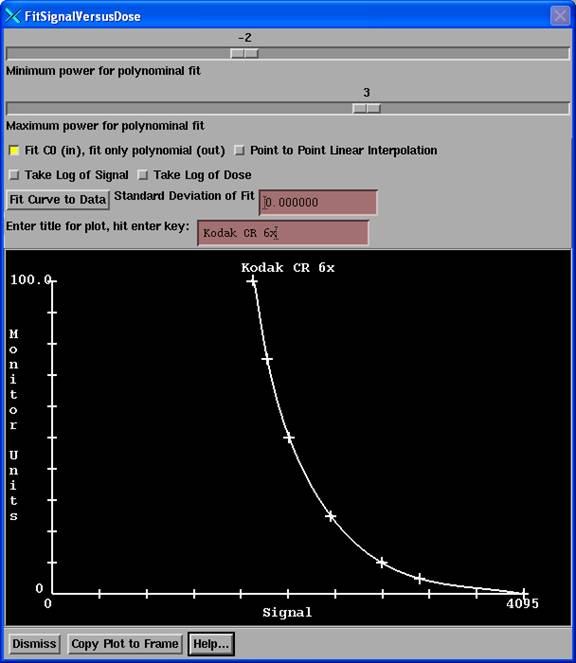
Note that above we fitted powers of (x-c) from –2 to +3 instead of powers of x. You may have to play with the range of powers to get a good curve fit. The toggle button “Fit C0” selects to fit powers of (x-c) instead of powers of x (or (c-x) if the signal were increasing with mu).
Using fit of (x-c) with powers –2 to +3 for this example we got back at the fit points:
|
Mu |
From Fit |
% error |
|
5 |
4.99 |
0.2 % |
|
10 |
10.00 |
0.0 % |
|
25 |
25.02 |
0.1 % |
|
50 |
50.02 |
0.0 % |
|
75 |
75.18 |
0.2 % |
|
100 |
100.01 |
0.0% |
Using log of mu versus signal resulted in errors as large as 2.8% as shown below:
|
Mu |
From fit |
% error |
|
5 |
5.05 |
1.0 % |
|
10 |
9.82 |
1.8 % |
|
25 |
24.52 |
2.0 % |
|
50 |
51.38 |
2.8 % |
|
75 |
76.13 |
1.5 % |
|
100 |
98.45 |
1.6 % |