
|
www.MathResolutions.com
Software Products for the Radiological Sciences
| Search |
|
www.MathResolutions.comSoftware Products for the Radiological Sciences |
|
| Home Page | Product Review | Program Manuals | Download Programs | Purchase | Site Map |
| Dosimetry Check | MarkRT (VGRT) | RtDosePlan | System 2100 | MillComp | C++ Library |
Here we will compare the results of a 3D dose reconstruction using the MapCheck diode array as an input device versus using the higher resolution Elekta EPID system. This case was supplied courtesy of the University of Toledo, Toledo, Ohio.
On the left is one of seven IMRT fields integrated with the MapCheck
diode array, and on the right is the same field integrated
with the Elekta EPID system.
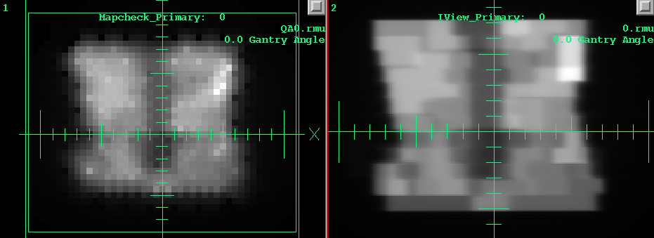
Although the MapCheck diode array has a much lower resolution, we must
remember that the patient acts like a low pass filter in regard
to the resulting dose distribution due to scatter within the patient.
Below is comparsion of the reconstructed dose on the left using the
MapCheck diode array, and on the right using the Elekta EPID,
for the transverse plane through isocenter. Green is the dose from the
planning system, magenta that reconstructed here.
Notice also the gamma analysis comparision for the same planes
using 3% - 3mm criteria, plotting the gamma value of 1.0.
Red is where the dose > plan dose, blue where dose < plan dose.
![]()
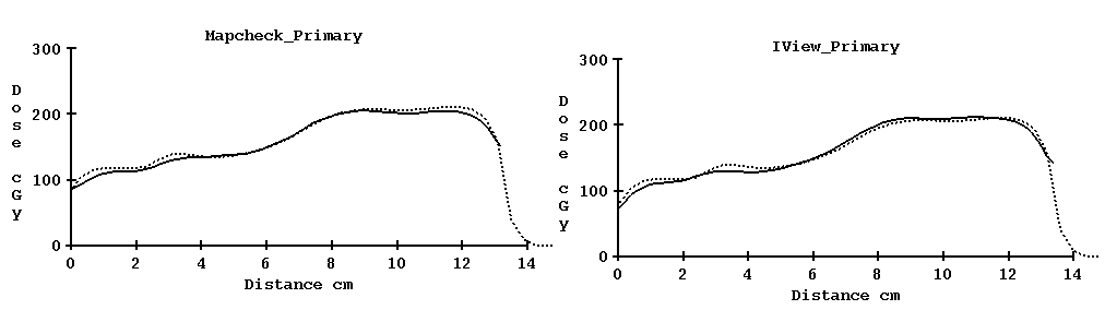
Next we show the profile through isocenter from patient posterior to anterior.
The dotted curve is from the planning system. On the left is
the profile from using the MapCheck as an input device, on the
right from using the Elekta EPID.
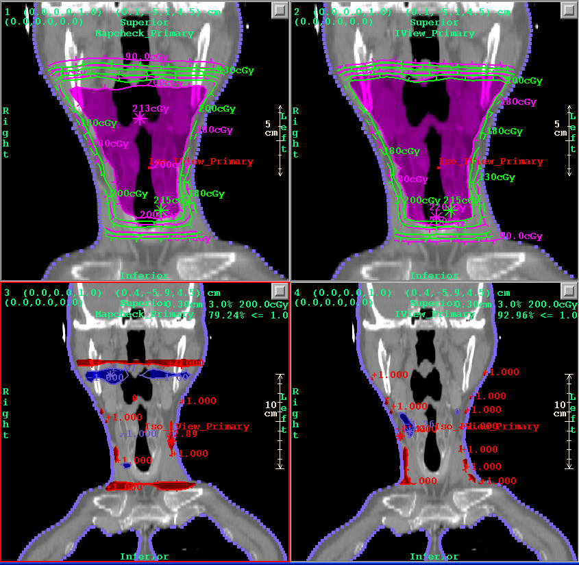
Below is comparsion of the reconstructed dose on the left using the
MapCheck, and on the right using the Elekta EPID,
for the coronal plane.
Notice also the gamma analysis comparision for the same plane
using 3% - 3mm criteria.
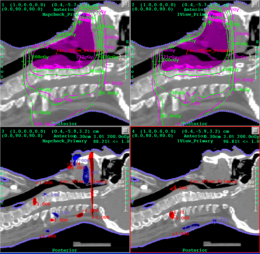
Below is comparsion of the reconstructed dose on the left using the
MapCheck, and on the right using the Elekta EPID,
for the sagittal plane.
Notice also the gamma analysis comparision for the same plane
using 3% - 3mm criteria.
Below is comparsion of the gamma volume histogram using the
MapCheck, and on the Elekta EPID, for 3%-3mm, for the patient
body volume.
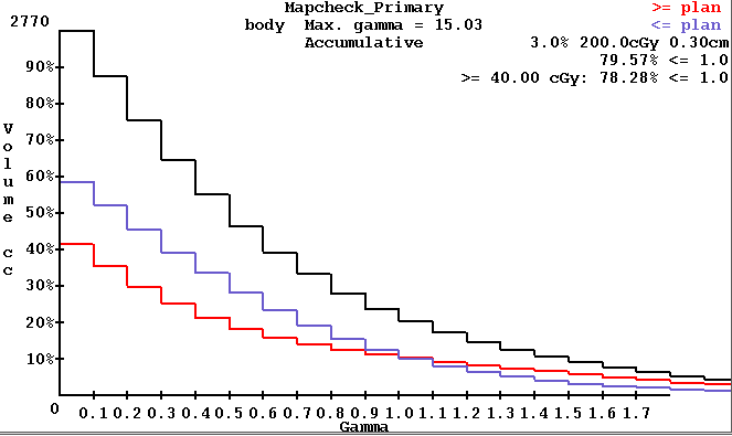
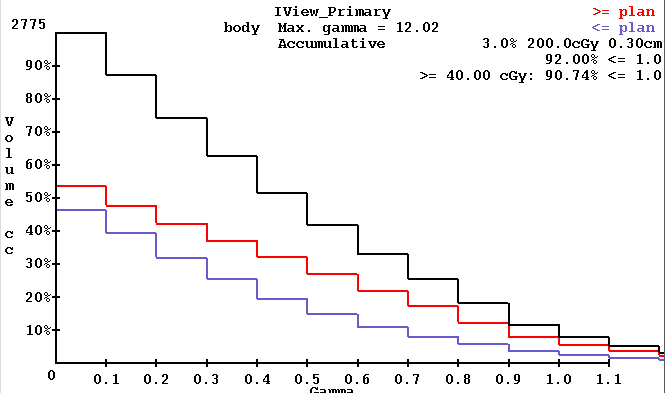
This example demonstrates that 3D dose reconstructedion can be done using the MapCheck diode array to integrate the radiation fields. There was some lose in the gamma volume histogram statistic, which in this case went from 92% to 80%.
Return to DosimetryCheck page
Return to homepage