
|
www.MathResolutions.com
Software Products for the Radiological Sciences
| Search |
|
www.MathResolutions.comSoftware Products for the Radiological Sciences |
|
| Home Page | Product Review | Program Manuals | Download Programs | Purchase | Site Map |
| Dosimetry Check | MarkRT (VGRT) | RtDosePlan | System 2100 | MillComp | C++ Library |
Here we will compare the results of a 3D dose reconstruction using the PTW 2D 729 ion chamber array versus using the higher resolution Varian EPID system. This case was supplied courtesy of Arthur Pinkerton of the North Mississippi Medical Center in Tupelo, Miss.
On the left is one of seven IMRT fields integrated with the PTW 2D
729 ion chamber array, and on the right is the same field integrated
with the Varian EPID system. In both cases an MLC carriage shift
required two separate integrations for each half.
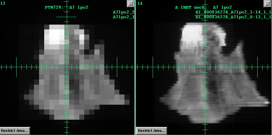
Although the PTW device has a much lower resolution, we must
remember that the patient acts like a low pass filter in regard
to the resulting dose distribution due to scatter within the patient.
Below is comparsion of the reconstructed dose on the left using the
PTW 2D ion chamber array, and on the right using the Varian EPID,
for a transverse plane.
Notice also the gamma analysis comparision for the same plane
using 3% - 3mm criteria.
![]()
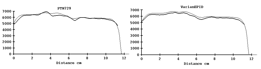
Next we show the profile through isocenter from patient right to left.
The dotted curve is from the planning system. On the left is
the profile from using the PTW 729 as an input device, on the
right from using the Varian EPID.
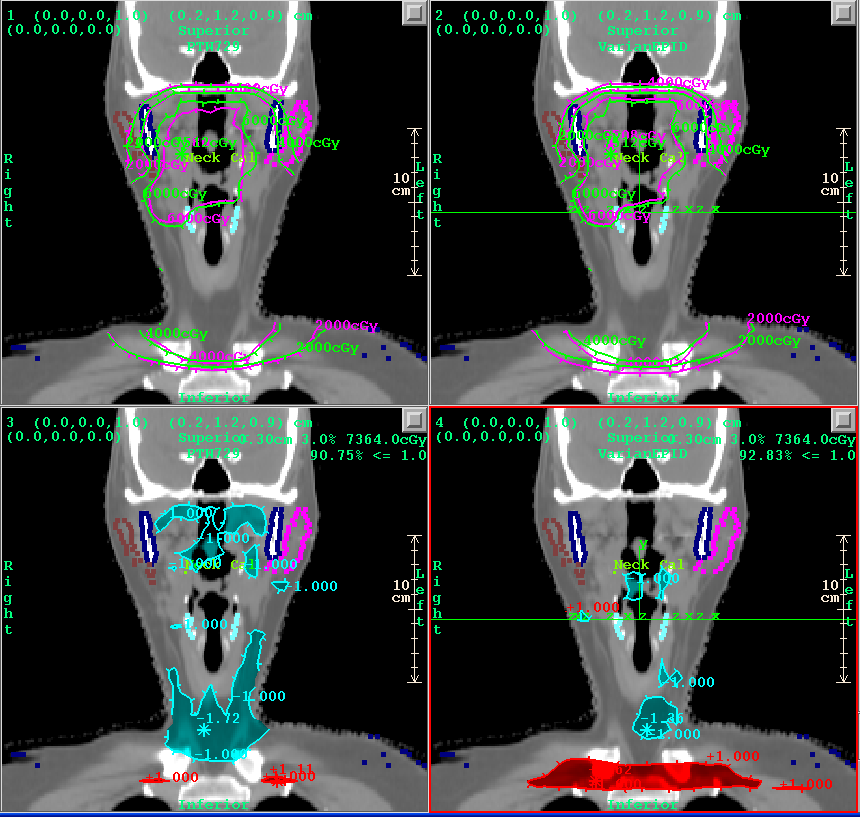
Below is comparsion of the reconstructed dose on the left using the
PTW 2D ion chamber array, and on the right using the Varian EPID,
for the coronal plane.
Notice also the gamma analysis comparision for the same plane
using 3% - 3mm criteria.
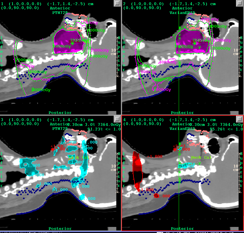
Below is comparsion of the reconstructed dose on the left using the
PTW 2D ion chamber array, and on the right using the Varian EPID,
for the sagittal plane.
Notice also the gamma analysis comparision for the same plane
using 3% - 3mm criteria.
Below is comparsion of the gamma volume histogram using the
PTW 2D ion chamber array, and on the Varian EPID,
for 3%-3mm.
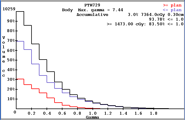
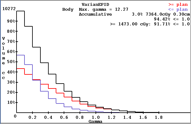
This example demonstrates that 3D dose reconstruction can be done using the PTW 2D 729 ion chamber array to integrate the radiation fields. There was no loss in the gamma volume histogram statistic in the example shown here.
Return to DosimetryCheck page
Return to homepage